LSCS/Cesarean
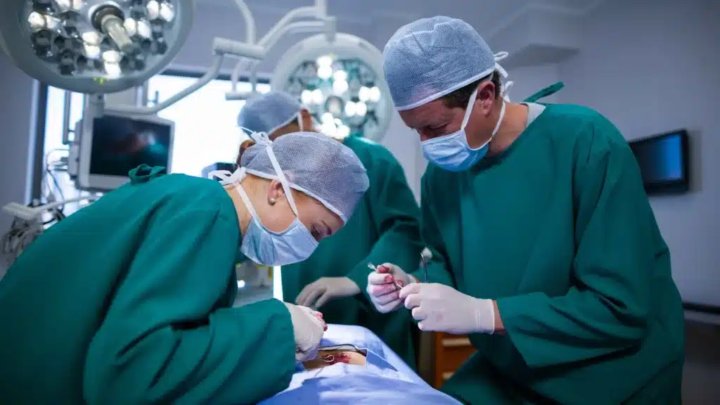
A Cesarean Section (C-section) is a surgical procedure performed to deliver a baby through an incision made in the abdomen and uterus. At Sahaj Hospital, we provide specialized care for both emergency and elective C-sections, ensuring the health and safety of both mother and baby. Our experienced obstetricians and surgeons are dedicated to offering safe, compassionate care for women undergoing this important procedure.
Our Specialties

What is a Cesarean Section?
A Cesarean Section (C-section) is typically performed when a vaginal delivery might pose a risk to the mother, the baby, or both. This may be due to various factors such as maternal health concerns, complications during labor, or fetal distress. In certain cases, a C-section is planned, either for medical reasons or at the request of the parents.
A Lower Segment Cesarean Section (LSCS), which is the most common type of C-section performed today, involves a transverse incision made just above the bladder. This technique is preferred because it minimizes blood loss and allows for faster recovery compared to other types of C-sections.
When is a Cesarean Section Necessary?
A C-section is often recommended in situations where a vaginal delivery would be too risky for the mother or baby. Some common indications for a C-section include:
Maternal Factors:
- Previous C-sections, especially classical (vertical) incisions
- Active genital herpes infection
- Cervical carcinoma (cancer of the cervix)
- Trauma to the mother, such as in an accident
- Maternal health conditions, such as high blood pressure or diabetes, that complicate labor
Fetal Factors:
- Cephalopelvic disproportion (the baby’s head is too large to pass through the birth canal)
- Placenta previa (when the placenta covers the cervix)
- Placental abruption (when the placenta detaches from the uterus prematurely)
- Fetal malposition, such as breech or transverse presentation
- Fetal distress (signs that the baby is not coping well during labor)
- Umbilical cord prolapse or cord compression
Complications During Labor:
- Failure of labor to progress, especially after attempted operative vaginal delivery
- Post-term pregnancy, where the baby has not been delivered by 42 weeks
At Sahaj Hospital, our team carefully evaluates all factors before recommending a C-section. Whether it’s for an emergency or a planned procedure, we ensure that the decision is made in the best interest of both mother and child.
The LSCS Procedure at Sahaj Hospital
Our expert obstetricians and surgical teams follow a precise, step-by-step process to ensure the safety of both mother and baby during an LSCS (Lower Segment Cesarean Section).
1. Anesthesia: Most C-sections are performed under spinal anesthesia, which numbs the lower body while allowing the mother to stay awake and alert during the procedure. In certain cases, general anesthesia may be required, especially if there are emergency situations like severe preeclampsia or eclampsia.
2. Preparing the Surgical Site: The abdomen is carefully cleaned and draped to minimize the risk of infection. Our team uses non-adhesive drapes and sterile techniques to protect both the mother and the surgical team.
3. Abdominal Incision: A horizontal incision is made just above the pubic area (Pfannenstiel incision). This type of incision is not only effective but also results in less postoperative pain and is cosmetically more acceptable to most patients.
4. Uterine Incision: The surgeon carefully opens the uterus, usually in a transverse direction, which is less invasive and leads to quicker recovery. The bladder is gently pushed aside, and the membranes around the baby are ruptured.
5. Delivery of the Baby: The baby is carefully lifted out of the uterus. In cases where the baby is in an unusual position, forceps or other instruments may be used to help with delivery.
6. After Delivery: The umbilical cord is clamped and cut, and the placenta is removed. The uterus is inspected to ensure that there are no remaining tissues or complications. Oxytocin may be administered to stimulate uterine contractions and reduce bleeding.
7. Closing the Incision: The uterine incision is closed in multiple layers to ensure that it heals properly. The abdominal muscles and skin are also closed with sutures that provide strength and support during the healing process.
Post-Operative Care and Recovery
After a Cesarean Section, the mother will typically stay in the hospital for 3-5 days. During this time, our healthcare team will monitor both the mother and baby for any signs of complications. Pain management, wound care, and support with breastfeeding are all essential components of the recovery process.
Most women who have had a C-section are able to resume normal activities within 6-8 weeks, although recovery times can vary depending on the individual. Our doctors provide detailed post-operative care instructions and support for a smooth recovery.
Potential Risks and Complications
Like any surgery, a Cesarean Section carries certain risks, although serious complications are rare. Some potential risks include:
During Surgery:
- Uterine atony (failure of the uterus to contract after delivery)
- Injury to surrounding organs, such as the bladder
- Heavy bleeding or hemorrhage
After Surgery:
- Wound infection or wound dehiscence (wound opening)
- Urinary tract infections
- Blood clots
- Abdominal wall hematoma (collection of blood in the abdominal wall)
At Sahaj Hospital, we take every precaution to minimize these risks and ensure that our patients receive the best care possible before, during, and after surgery.
