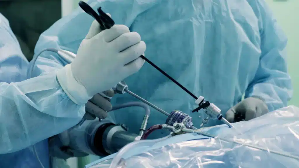
Laparoscopic Myomectomy
Welcome to Sahaj Hospital, Indore, home to MP’s first and only dedicated Fibroid Clinic. Our specialized Laparoscopic Myomectomy services provide advanced, minimally invasive solutions for the treatment of uterine fibroids. With cutting-edge technology, expert surgeons, and a patient-centered approach, we aim to deliver the highest quality care tailored to your unique needs.
For reliable and effective Laparoscopic Myomectomy in Indore, you can trust our expertise. Our commitment to excellence and compassionate care makes us the leading choice for uterine fibroid treatment. Book your consultation now and take the first step toward a healthier, symptom-free life.
Our Specialties

Understanding Uterine Fibroids
Uterine fibroids, also known as leiomyomas or uterine myomas, are non-cancerous growths that develop from the muscular tissue of the uterus (womb). These growths can range in size from a tiny seed to as large as a melon and may occur as a single fibroid or multiple fibroids. Although fibroids are benign, they can lead to uncomfortable symptoms such as heavy menstrual bleeding, pelvic pain, and complications in fertility or pregnancy.
Types of Uterine Fibroids:
- Intramural Fibroids: Develop within the wall of the uterus; the most common type.
- Subserosal Fibroids: Form on the outer wall of the uterus and may grow on stalks, becoming pedunculated.
- Submucosal Fibroids: Located beneath the lining of the uterine wall, often causing significant symptoms.
- Cervical Fibroids: Found in the cervix, the lower part of the uterus.
Symptoms of Uterine Fibroids
While some women experience no symptoms, others may face challenges such as:
- Heavy, painful menstrual periods
- Lower abdominal swelling
- Frequent urination
- Backache or leg pain
- Pain during sexual intercourse
- Infertility or recurrent miscarriages
Diagnosis of Uterine Fibroids
Fibroids are often diagnosed during a routine pelvic examination or through imaging techniques such as:
- Ultrasound: The most common method to detect fibroids and rule out other conditions.
- Transvaginal Ultrasound: Provides a detailed view by inserting a scanner into the vaginal canal.
- Hysteroscopy: A telescope-like device to examine the uterus lining and obtain tissue samples if necessary.
- Laparoscopy: A minimally invasive technique to assess the fibroids’ size, number, and location.
- Biopsy: To rule out rare malignant growths like leiomyosarcoma.
How is Laparoscopic Myomectomy Performed?
During laparoscopic myomectomy:
- Small incisions are made near the navel.
- A laparoscope, equipped with a camera, provides a clear view of the internal organs.
- Special surgical instruments are used to remove the fibroids.
- The fibroids are fragmented and removed through small openings.
- The uterus is reconstructed to preserve its normal function.
This advanced technique is effective for treating intramural, subserosal, and even complex fibroids, making it a preferred choice for women looking to resolve symptoms and maintain reproductive potential.
Recovery and Follow-Up
Most patients resume normal activities within a few weeks post-surgery. Our team emphasizes follow-up care to monitor healing and address any concerns. Regular check-ups help maintain uterine health and improve fertility outcomes.

