What happens to the joint in osteoarthritis?

Osteoarthritis is the commonest form of arthritis. Most affected is the knee joint. It is a degenerative disease in which “wear-and-tear” happens. Age more than 50 years are more likely to get this disease, but nowadays even young people are presenting with this disease.

There is a protective tissue between the bones that help in smooth movements of the bones. Degradation causes cartilage damage and hampers the smooth movement of the bones. After complete degeneration of the cartilage, the bone-ends start rubbing against each other causing severe pain and spurring of the bones.

This disease develops gradually and with degeneration, the pain worsens.

How to diagnose osteoarthritis?

  • X-ray (radiograph): X-ray of the knee taken in the standing position, helps in cartilage damage assessment.
  • Computed tomography scan (CT or CAT scan): This radiological modality gives a better perception of the damage than X-ray alone.
  • Magnetic resonance imaging (MRI): Skeletal structures, blood vessels, and tissues can be visualized properly with this technique.
  • Arthroscopy: This procedure which is minimal access shows the inside of the knee joint and also does procedures to correct the errors. This procedure requires anesthesia.

What is joint preservation?

It is a novel treatment modality that emphasizes pain relief using biological mechanisms.

What are the types?

Nonsurgical treatment

  • Treatment with Autologous Adipose-Derived Stromal Vascular Fraction

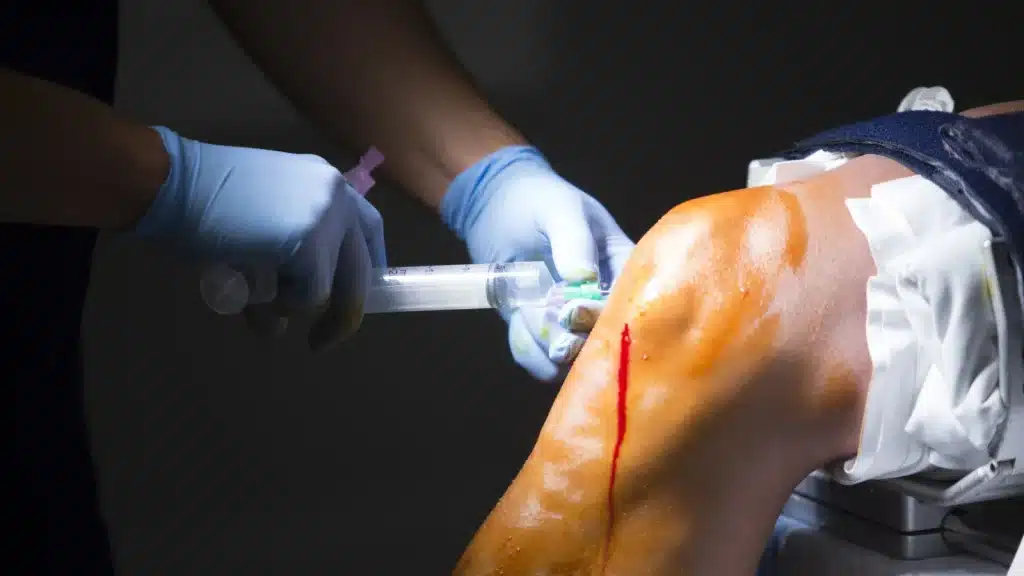
    Adipose-derived Stromal Vascular Fraction (Ad-SVF) contains a heterogeneous mix of adipose stromal cells, (progenitor cells, pericytes, T-regulatory cells, endothelial progenitor cells, anti-inflammatory M2 macrophages, extracellular matrix, and many other cells types.) The articular space receives these cells through an injection into the joint space. These cells then attach to the areas of damage and form/repair these areas. After anchoring to the damaged tissue, these cells form the same tissue.

    Thus, they help in the formation of the cartilage and repair of the other tissues inside the joint.

    • The process is done in these basic steps –
      • Lipoaspiration of adipose tissue,
      • Ultrasonic cavitation, centrifugation,
      • And filtration takes around 40 minutes.
    • The stromal vascular fraction is very safe in comparison to other treatment modalities
      • It is autologous, i.e. cells are from the same person
      • The processing and administration of the injection are done in the same single surgical sitting
      • There are no age limitations for this treatment
      • Preferred in the early grade of osteoarthritis
      • BMI of the patient is not a limiting factor
      • Done under anesthesia
      • Patients can start walking from the same day
      • The intraarticular space receives the stromal vascular fraction (i.e. within the articular space of the joint).
      • The adipose tissue obtained is not cultured using any enzymes or chemical or animal product.
      • The patient can feel the effect (reduction in pain) within the first week of the treatment and by 3-4 weeks, the patient can get back to their normal activities.
      • There is no need for bed rest or any other external support.
      • There is clinical evidence that reported the growth of cartilage inside the knee joint over time. Thus, it provides long-term benefits and in many cases, there is no requirement for any further intervention.
    • This therapy is not recommended in the following patients:
      • If the patient is pregnant,
      • Having any blood disorders
      • Too lean to obtain sufficient adipose tissue
      • Having any ongoing infection
      • Having gout or pseudogout
      • Having any chronic terminal disease like cancer
    • But if there is severe deterioration of knee cartilage (late-stage osteoarthritis), the only option available is to go for surgical intervention.
Share via
Copy link
Powered by Social Snap